

 |
 |
 |
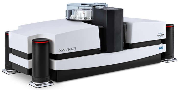
The desktop SKYSCAN 1272 CMOS is an innovative high-resolution 3D X-ray microscope based on micro computed tomography (micro-CT) technology, and builds on the trusted SKYSCAN 1272 platform to integrate the latest X-ray technologies.
Its state-of-art 16 megapixel sCMOS X-ray detector provides high-contrast images with superior resolution. The extended detector field of view and enhanced sensitivity for X-rays result in up to twice shorter scan times. The extraordinary native resolution of up to 11200 x 11200 pixels per slice allows zooming into any part of the 3D volume without rescanning the sample. The new Clean ImageTM scan mode significantly reduces typical CT artefacts right from the start, thus providing great quality images without cumbersome posteriori corrections.
This top performance is paired with low cost of ownership. Our desktop SKYSCAN 1272 CMOS Edition can be placed on any laboratory desk and consequently does not occupy a lot of expensive lab space. A standard domestic power plug is all you need to start running the instrument, no water chiller or additional compressor. There are also no further hidden costs as the industry-grade sealed X-ray source is maintenance-free. SKYSCAN 1272 is complemented by 3D.SUITE. This comprehensive software suite covers GPU-accelerated reconstruction, 2D/ 3D morphological analysis, as well as surface and volume rendering visualization.
The SKYSCAN 1272 offers automatic selection of parameters with Genius-Mode. Magnification, energy, filter, exposure time and background corrections can all be optimized automatically with a single click.
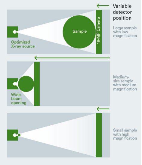
Furthermore, both the sample and large-format sCMOS camera can be positioned as close as possible to the source, which substantially increases the measured intensity. That is why SKYSCAN 1272 scans up to 5 times faster compared to conventional systems with fixed camera position.
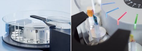
SKYSCAN 1272 can optionally be equipped with an external 16-position sample changer to increase throughput for QC and routine analysis.
The sample changer accepts a variety of sample sizes, up to a diameter of 25 mm.
Samples can be can be easily replaced at any time without interrupting an ongoing scanning process. New samples are automatically detected, and LED‘s indicate the status for every scan: ready, scanning, done.
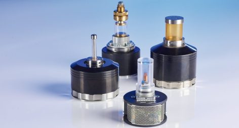
The Bruker material testing stages are designed to perform compression experiments up to 4400 N and tensile experiments up to 440 N. All stages automatically communicate through the system’s rotation stage, without the need of any cable connections. Using the supplied software, scheduled scanning experiments can be set up.
Bruker's heating and cooling stages can reach temperatures of up to +80ºC, or 30ºC below ambient temperature. Just like the other stages, no extra connections are needed, and there is an automatic recognition of the stage. Using the heating & cooling stages, samples can be examined under non-ambient conditions, to evaluate the effect of temperature on the sample’s microstructure.
The SKYSCAN 1272 succeeds the SKYSCAN 1072 and SKYSCAN 1172 models, between them accounting for at least half of the entire microCT bone morphometry literature. It’s the benchmark for high resolution with high throughput and ease of use. This makes it the ideal solution for bone disease models from osteoporosis and arthritis to bone tumor and myeloma, and for large scale gene phenotyping.
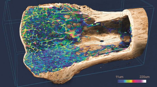
Ex vivo scanning of biological tissues is a very good way to show their internal structures non-destructively. A contrast agent or chemical drying can improve the image quality by further enhancing or differentiating densities. The SKYSCAN 1272 is the ideal platform for such imaging with its resolution, easy sample handling and high throughput – that’s why you will find so many publications using this scanner.
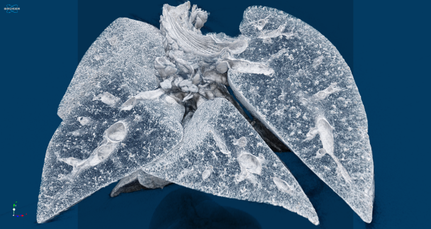
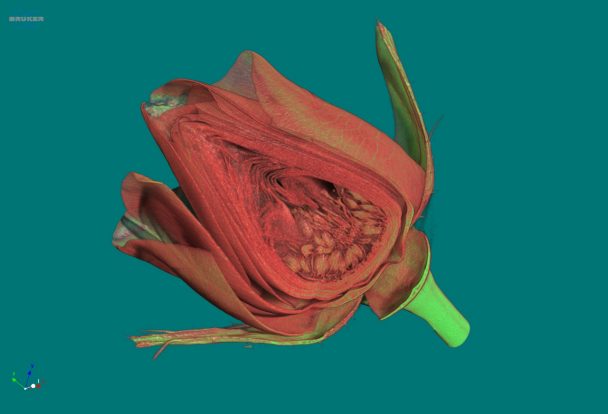
MicroCT is exceptionally good for the visualization of internal structures in the finest details of the tissues of plants and animals. This imaging method differentiates between densities without harming or destroying the scanned object. That’s why zoology and botany are fast-growing microCT applications with SKYSCAN 1272 users at the forefront. A wide range of different living organisms can be visualized and analyzed with minimal or no sample-treatment.


|
|
|